Now that I've covered the basic function of antibodies and how they're made, I think I'll turn my attention to immobilization techniques. There are a number of different ways to stick antibodies onto a solid surface, and the strategy that is used is mostly dependent on the type of surface you're working with. Gold, for example, is pretty easy. This is because the cystein residues that are present all over the antibody structure will bind - albeit fairly weakly - to gold. There is a natural attraction between the thiol group of the cystein residue and the gold. So when you expose IgG antibodies to gold at a nice comfortable pH of 7.5-8.5, the antibodies will adsorb onto the gold surface. Although this method is easy and relatively effective, it does not create a very stable bond.
To immobilize antibodies with a stronger binding scheme, the protein must be covalently attached to the surface. This type of covalent linkage between a surface and a protein is often used when the surface is glass. Glass is a material whose surface is surprisingly easy to modify using a class of compounds called silanes. Silane molecules are most often based around a single silicon atom. The silicon atom has three ethoxy or methoxy groups. These groups will covalently bind to glass, creating extremely stable bonds that are also able to crosslink with other nearby silanes to further stabilize the silane layer. The fourth valence electron is bound to an organic species - usually a functional group connected to the silicon via a short hydrocarbon linker. One of the more common silanes used in immobilization techniques is mercaptopropyl(triethoxysilane), and its structure looks like this:
By looking at the structure, you can clearly see the three ethoxy groups (O-CH3) bound directly to the Si atom, and the one mercapto group (SH) connected to the Si atom by a three-carbons (propyl) bridge.
So once the surface of the glass is functionalized with a silane layer, it is much more reactive than the fairly inert native glass surface. The next step would then be to connect the functional layer of the modified glass surface to one of the amino acids of the IgG antibody. This is accomplished through the use of a crosslinker. To give give an example, the crosslinker that I have the most experience with is GMBS (long chemical IUPAC name: 4-Maleimidobutyric acid N-hydroxysuccinimide ester). And this is what it looks like:
GMBS is known as a heterobifunctional crosslinker because the two ends of the molecule are different and are reactive towards different type of functional groups. In this case, the maleimide group on the left binds covalently to the mercapto group of the functionalized glass. The succinimidyl ester on the right then binds to amine groups found on the peptide chains that make up the antibody protein. Once this reaction successfully completes - which happens fairly quickly - you end up with a glass surface that is coated in IgG antibodies. And therefore, the surface is now capable of selectively binding the antigen of interest.
One last note about antibody immobilization: the the steric position of the antibodies on the glass is important. By simply crosslinking the protein directly to the glass surface, you have no way of controlling the position of the antibody. For instance, the crosslinkage could occur at or near the antigen binding site of antibody. This would mean that this particular antibody would end up immobilized 'upside down,' with the antigen binding sites so close to the glass surface that the antigen would be unable to bind. To remediate this problem, you can first crosslink special proteins, such as Protein A or Protein G, to the surface. Protein A and Protein G have a binding site that is specific to a highly conserved region near the 'bottom' of the antibody, on the opposite side of the IgG from the antigen binding sites. After immobilization of Protein A or G, you can introduce the antibody, it will bind to the Protein A or G, and you end up with a surface in which all of the antibodies are pointed 'up' with the antigen binding sites exposed and available.
Showing posts with label Biosensors. Show all posts
Monday, September 19, 2011
Wednesday, September 14, 2011
A Few Notes on Antibodies: Part 1
During my doctorate research, I shifted my research focus from more traditional biosensor technologies, such as immunosensors, to sensing applications of molecularly imprinted polymers. However, I still dabble in immunosensors fairly often, and my expertise and previous experience in antibody-based sensors comes in handy, particularly for other researchers who come to me with questions and guidance in this field. Here at LU, my research group is currently waiting to receive a fluorescent microscope that we recently ordered. When the new scope arrives, I will be using it to conduct a study examining targeted antibody immobilization onto gold electrodes. The plan is to block specific areas of our sensor substrate so that antibodies will only be able to bind to certain regions. When fluorescent bacteria are introduced, they'll bind specifically to the antibodies, and this phenomenon should be clearly visible under the fluorescent microscope. So in the spirit of beginning this work, I thought I'd discuss some of the basics of antibodies and antibody immobilization. I'm going to be brief, so I may have to turn this into a series of posts.
First off, I need to clarify one thing: when I say antibody, I am referring solely to immunoglobulin G (IgG) antibodies. Next, I guess I should describe what antibodies are and what they do. The IgG antibody is a relatively high molecular weight protein. It's produced by our immune cells (B cells) in response to infection. When the antibodies are produced during infection, they bind to the bacterium or virus or whatever, coating its surface and acting as a sort of signaling beacon. Other immune cells are able to detect this beacon and attack and destroy the infection. So in a very general sense, that's the natural function of antibodies.
This natural function of the IgG antibody is made possible by one of its most important properties; selectivity. The IgG antibody is a large Y-shaped protein that looks something like this:
At the top ends of the two 'arms' of the antibody are binding sites that are capable of binding to one single antigen, which is the target bacteria or virus or whatever, and only that one antigen. This is what is referred to as selectivity.
Researchers like me who are interested in using the properties of antibodies for biosensors and other applications must be able to readily produce or purchase these antibodies. As it turns out, there is a fairly effective way of harvesting the antibodies that are selective for a particular antigen of interest. Let's say, for instance, that you want an antibody that is selective for the flu virus. To produce these antibodies, you would take a syringe that contained the flu virus and inject it into the lumen of the gut - or some other space where the antigen would not leak out into the bloodstream - of a mammal. The mammal used is commonly a mouse, rabbit, or goat. The B cells infiltrate the gut lumen of the animal and begin churning out antibodies that are selective for the flu virus. Then you would go back and draw out the fluid from the lumen of the gut and purify the antibodies that were produced. And there you have it - anti-influenza antibodies.
Having only touched the surface of this topic, there will definitely be more to come.
First off, I need to clarify one thing: when I say antibody, I am referring solely to immunoglobulin G (IgG) antibodies. Next, I guess I should describe what antibodies are and what they do. The IgG antibody is a relatively high molecular weight protein. It's produced by our immune cells (B cells) in response to infection. When the antibodies are produced during infection, they bind to the bacterium or virus or whatever, coating its surface and acting as a sort of signaling beacon. Other immune cells are able to detect this beacon and attack and destroy the infection. So in a very general sense, that's the natural function of antibodies.
This natural function of the IgG antibody is made possible by one of its most important properties; selectivity. The IgG antibody is a large Y-shaped protein that looks something like this:
At the top ends of the two 'arms' of the antibody are binding sites that are capable of binding to one single antigen, which is the target bacteria or virus or whatever, and only that one antigen. This is what is referred to as selectivity.
Researchers like me who are interested in using the properties of antibodies for biosensors and other applications must be able to readily produce or purchase these antibodies. As it turns out, there is a fairly effective way of harvesting the antibodies that are selective for a particular antigen of interest. Let's say, for instance, that you want an antibody that is selective for the flu virus. To produce these antibodies, you would take a syringe that contained the flu virus and inject it into the lumen of the gut - or some other space where the antigen would not leak out into the bloodstream - of a mammal. The mammal used is commonly a mouse, rabbit, or goat. The B cells infiltrate the gut lumen of the animal and begin churning out antibodies that are selective for the flu virus. Then you would go back and draw out the fluid from the lumen of the gut and purify the antibodies that were produced. And there you have it - anti-influenza antibodies.
Having only touched the surface of this topic, there will definitely be more to come.
Labels:
antibodies,
Biosensors,
immunoglobulin g,
immunosensors
Tuesday, April 26, 2011
Shine On You Deadly Bacteria
As I mentioned in a previous post, I've been working with a small research team on the development of an electrochemical sensor to detect pathogenic E. coli bacteria. I've already discussed the difficulty that I had in obtaining an accurate cell count to quantify the concentration of the E. coli cells in the soy broth suspension that they live in. Well, I've overcome the problems that I was having with cell counting and have moved on to fluorescent imaging of the cells. More specifically, I've been charged with labeling the E. coli with a fluorescent dye and exposing the labeled cells to our electrochemical devices functionalized with IgG antibodies that selectively bind to E. coli. Then, rather than taking electrochemical measurements, I want to take fluorescent images to correlate our findings from the sensor data to actual images of the cells bound to the device.
I've been working on fluorescently labeling the E. coli cells with fluorescein isothiocyanate (FITC) for a couple of months now, with essentially no success. FITC is an amine-reactive fluorophore, and so my plan was to label the anti-E. coli antibody with FITC, then allow the labeled antibodies to coat the surface of the cells. However, that didn't work out so well. After several attempts using this procedure, I ended up with zero observable or measurable fluorescence from the E. coli cells. I did learn some things along the way, though. Most notably, I learned that trying to label viable live cells is a fool's errand unless absolutely necessary, so I started heat killing the cells before the labeling procedure.
After these failed attempts, I decided to simplify things a bit. Why not try to label the E. coli with FITC directly, rather than trying to use the antibody as an intermediate? So that's what I did. I introduced the killed cells to a large concentration of FITC, allowed the binding reaction to take place for an hour or so, and then purified the labeled cells from the remaining free dye. The resulting solution of labeled E. coli showed strong fluorescence when analyzed using fluorescence spectroscopy, so I dropped the cell suspension on a microscope slide and took some fluorescent images. And...well...see for yourself:

Those small elongated dots are the brightly labeled rod-shaped bacteria. I should mention that, despite being stored in the dark, the fluorescent emission of the cells is dropping surprisingly quickly. I would guess that this may be an indication that rather than the expected covalent attachment of the FITC to surface proteins on the E. coli, the FITC diffused into the cell and is slowly diffusing back out again over time. Either way, the cells were initially nice and bright, which allowed me to take some good images of the labeled cells bound to our electrochemical sensor device, which is exactly what I needed to do.
I've been working on fluorescently labeling the E. coli cells with fluorescein isothiocyanate (FITC) for a couple of months now, with essentially no success. FITC is an amine-reactive fluorophore, and so my plan was to label the anti-E. coli antibody with FITC, then allow the labeled antibodies to coat the surface of the cells. However, that didn't work out so well. After several attempts using this procedure, I ended up with zero observable or measurable fluorescence from the E. coli cells. I did learn some things along the way, though. Most notably, I learned that trying to label viable live cells is a fool's errand unless absolutely necessary, so I started heat killing the cells before the labeling procedure.
After these failed attempts, I decided to simplify things a bit. Why not try to label the E. coli with FITC directly, rather than trying to use the antibody as an intermediate? So that's what I did. I introduced the killed cells to a large concentration of FITC, allowed the binding reaction to take place for an hour or so, and then purified the labeled cells from the remaining free dye. The resulting solution of labeled E. coli showed strong fluorescence when analyzed using fluorescence spectroscopy, so I dropped the cell suspension on a microscope slide and took some fluorescent images. And...well...see for yourself:

Those small elongated dots are the brightly labeled rod-shaped bacteria. I should mention that, despite being stored in the dark, the fluorescent emission of the cells is dropping surprisingly quickly. I would guess that this may be an indication that rather than the expected covalent attachment of the FITC to surface proteins on the E. coli, the FITC diffused into the cell and is slowly diffusing back out again over time. Either way, the cells were initially nice and bright, which allowed me to take some good images of the labeled cells bound to our electrochemical sensor device, which is exactly what I needed to do.
Monday, November 22, 2010
Shmorgisborg of Science
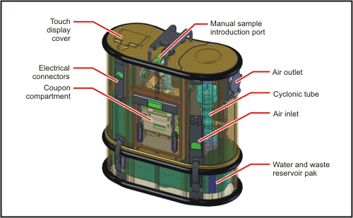
During my dissertation defense presentation a couple of weeks ago, I explained that one of the advantages of any fluorescence-based biological or chemical detection system would be the ability to scale down such a system to a portable and deployable size. My evidence for this was the mention of two biosensing detection systems commercialized by Research International: the Raptor and the Biohawk. Both of these systems are, for lack of a better word, awesome. Both systems use what are known as sandwich immunoassays to detect the target analytes. And both systems are capable of detecting multiple analytes simultaneously. The sandwich immunoassay used in these sensors is an antibody-based detection method in which a primary antibody specific to a target compound is adsorbed onto a glass waveguide, similar to an optical fiber. If the target compound is present in a sample, it will bind selectively to the primary antibody. After exposure to a sample, a solution containing a secondary antibody that is labeled with a fluorescent dye is flown over the waveguide. If the target compound is bound to the primary antibody, the secondary antibody will also bind to it, creating a sort of antibody-antigen-antibody sandwich (hence the name of the assay). Excitation light is then sent through the waveguide, exciting the fluorescent dye bound to the secondary antibody, indicating the presence of the target compound.
I remember learning about RI's Raptor sensor during my coursework early in my graduate studies, so it has been around for quite some time and has become a bit of a mainstay. Certainly if you'd like to learn more about either of these systems, you can check out Research International's website and read up about them. Besides plenty of reading material, there are some really nice diagrams on how the sandwich immunoassay works, as well as diagrams showing the inner workings of a fully automated biosensor. They also have some other cool detection and sampling platforms that are worth checking out if you're interested in those types of things.
And now for some news.
As if CERN hadn't made headlines enough lately (see Large Hadron Collider), they made big news last week when they announced and published the results of their recent studies on antimatter. One of two research groups working on parallel studies was able to effectively create atoms of antihydrogen, hold them in an electromagnetic field, and keep them there for nearly 200 milliseconds. Although this is a big deal, the end goal is to create and trap many more antihydrogens and to hold them for much longer periods of time so that the properties of the trapped antimatter can be thoroughly studied and examined. And so this study is a major step forward in regard to the overall objective.
And there you have it, folks. A blog update with actual substance. Been a while.
Wednesday, January 6, 2010
The Triumphant Return of the Cellular Biosensor
Cell-based biosensors kind of get a bad rap. And the reason for this is pretty simple: cells die. They're delicate little creatures that have to be continually monitored and cared for. And, when used as the basis of a sensing technique, if your cells die, your sensor doesn't work. That's why most sensing schemes use more hearty methods, such as antibodies or synthetic receptors or chemical reactions. This is the reason that I was surprised to see a cell-based sensor grabbing lots of attention in the media recently. In fact, I picked up the story initially from BBC news, but found via a quick internet search that the story had been published on news websites ranging from Science Daily to the India Report.
At any rate, the rationale behind the study was to examine the way in which a popular drug for treatment of schizophrenia works. It is known that the drug causes an increase in production of acetylcholine, but it had also been shown to block the receptors for acetylcholine. Without knowing which dichotomous action was prevailing within the interior of cells, it was impossible to deduce the action of the drug on the brain.
The research team then devised a cell-based biosensor to study the exact effect that the drug had on cells in the brain. The group began with embryonic kidney cells and genetically modified them so that the receptor for acetylcholine was directly coupled with a common G-protein downsteam intracellular signaling cascade. This cascade was then linked to calcium ion upregulation, which activated a calcium-sensitive fluorescent reporter. The fluorescent reporter, then, was fluorescence resonance energy transfer (FRET)-based, meaning that it consisted of two distinct fluorescent molecules. When exposed to calcium, the two fluorescent molecules move closer together, causing one of the molecules to donate more energy to the other, producing a visible color change. This means that when the cells bind acetylcholine, they produce calcium, which causes the color of their fluorescence to shift. When these cells were implanted into rat brains and the rats exposed to the schizophrenia drug, they found no change in the fluorescence of the sensor cells, meaning that the receptor blocking activity of the drug was its primary function.
At least, all this is what I gathered from their most recent publication in Nature. There you can take a look at the team's data as well as a more detailed description of their methods. Really, this is pretty exciting stuff. Utilizing cellular biosensors in this way could be a major player in future drug discovery research. No more guessing as to the function of drugs based on studies on cells in a petri dish. Implant the genetically modified sensor cells, give the rat the drug, and visually see the effect that the drug has on the cells.
At any rate, the rationale behind the study was to examine the way in which a popular drug for treatment of schizophrenia works. It is known that the drug causes an increase in production of acetylcholine, but it had also been shown to block the receptors for acetylcholine. Without knowing which dichotomous action was prevailing within the interior of cells, it was impossible to deduce the action of the drug on the brain.
The research team then devised a cell-based biosensor to study the exact effect that the drug had on cells in the brain. The group began with embryonic kidney cells and genetically modified them so that the receptor for acetylcholine was directly coupled with a common G-protein downsteam intracellular signaling cascade. This cascade was then linked to calcium ion upregulation, which activated a calcium-sensitive fluorescent reporter. The fluorescent reporter, then, was fluorescence resonance energy transfer (FRET)-based, meaning that it consisted of two distinct fluorescent molecules. When exposed to calcium, the two fluorescent molecules move closer together, causing one of the molecules to donate more energy to the other, producing a visible color change. This means that when the cells bind acetylcholine, they produce calcium, which causes the color of their fluorescence to shift. When these cells were implanted into rat brains and the rats exposed to the schizophrenia drug, they found no change in the fluorescence of the sensor cells, meaning that the receptor blocking activity of the drug was its primary function.
At least, all this is what I gathered from their most recent publication in Nature. There you can take a look at the team's data as well as a more detailed description of their methods. Really, this is pretty exciting stuff. Utilizing cellular biosensors in this way could be a major player in future drug discovery research. No more guessing as to the function of drugs based on studies on cells in a petri dish. Implant the genetically modified sensor cells, give the rat the drug, and visually see the effect that the drug has on the cells.
Subscribe to:
Posts (Atom)