Showing posts with label escherichia coli. Show all posts

Monday, June 13, 2011

MUG: It's Not Just For Coffee Anymore


Recently, I have been playing around with a special cell culture media that uses a fluorescent indicator to signal the presence of E. coli bacteria. The culture media is a nutrient broth called LST-MUG, and is often referred to by its proprietary name, Hach media. The nutrient broth is similar to other types of cell culture media, with a couple of notable exceptions. First, the media contains surfactants that inhibit the growth of non-coliform organisms. This basically means that everything but E. coli is killed in the broth, allowing only E. coli to grow in the media. Second, the media contains a molecule called 4-methylumbelliferyl-β-D-glucuronide, or MUG. MUG is the part of the media that is responsible for indicating the presence of E. coli.

Under normal conditions, MUG emits very weak fluorescence at a wavelength down around where blue and UV meet, somewhere around 400 nm, when exposed to UV light. When MUG is introduced to E. coli, it is cleaved (or cut) into two separate smaller molecules by an enzyme within the bacteria called
β-glucuronidase. One of the products of this cleavage reaction (the glucoronide part) is basically inert and does nothing at all. But the other product of the reaction is 4-methylumbelliferone, a highly fluorescent molecule. When exposed to the same UV light as before, the fluorescence of 4-methylumbelliferone is easily observable as a pale blue glow. In this way, the MUG acts a fluorogenic reporter of the presence of E. coli bacteria.

This bacterial growth media with the fluorescent reporter is commercially available and widely used to detect the presence of E. coli. However, it has some pitfalls. One problem with the media is that the sample to be tested must be introduced to the broth and then incubated, which is best done in a laboratory environment by a trained technician. A more pressing issue with the broth is its response time, which is 16-24 hours. If you are testing a sample of, say, raw beef, you would take a random in-line sample during processing and inoculate the Hach media. Twenty four hours later, a fluorescent signal is observed in the media, indicating that the beef is contaminated with E. coli. But that particular beef product is already on a truck on its way to the grocery store.

One of the projects that we're focusing on at LU's Center for Nanotechnology & Biosensors is a method for making this process much more effective and drastically faster, so that E. coli can be detected efficiently and contaminated products can be kept off of store shelves.

Tuesday, April 26, 2011

Shine On You Deadly Bacteria

As I mentioned in a previous post, I've been working with a small research team on the development of an electrochemical sensor to detect pathogenic E. coli bacteria. I've already discussed the difficulty that I had in obtaining an accurate cell count to quantify the concentration of the E. coli cells in the soy broth suspension that they live in. Well, I've overcome the problems that I was having with cell counting and have moved on to fluorescent imaging of the cells. More specifically, I've been charged with labeling the E. coli with a fluorescent dye and exposing the labeled cells to our electrochemical devices functionalized with IgG antibodies that selectively bind to E. coli. Then, rather than taking electrochemical measurements, I want to take fluorescent images to correlate our findings from the sensor data to actual images of the cells bound to the device.

I've been working on fluorescently labeling the E. coli cells with fluorescein isothiocyanate (FITC) for a couple of months now, with essentially no success. FITC is an amine-reactive fluorophore, and so my plan was to label the anti-E. coli antibody with FITC, then allow the labeled antibodies to coat the surface of the cells. However, that didn't work out so well. After several attempts using this procedure, I ended up with zero observable or measurable fluorescence from the E. coli cells. I did learn some things along the way, though. Most notably, I learned that trying to label viable live cells is a fool's errand unless absolutely necessary, so I started heat killing the cells before the labeling procedure.

After these failed attempts, I decided to simplify things a bit. Why not try to label the E. coli with FITC directly, rather than trying to use the antibody as an intermediate? So that's what I did. I introduced the killed cells to a large concentration of FITC, allowed the binding reaction to take place for an hour or so, and then purified the labeled cells from the remaining free dye. The resulting solution of labeled E. coli showed strong fluorescence when analyzed using fluorescence spectroscopy, so I dropped the cell suspension on a microscope slide and took some fluorescent images. And...well...see for yourself:


Those small elongated dots are the brightly labeled rod-shaped bacteria. I should mention that, despite being stored in the dark, the fluorescent emission of the cells is dropping surprisingly quickly. I would guess that this may be an indication that rather than the expected covalent attachment of the FITC to surface proteins on the E. coli, the FITC diffused into the cell and is slowly diffusing back out again over time. Either way, the cells were initially nice and bright, which allowed me to take some good images of the labeled cells bound to our electrochemical sensor device, which is exactly what I needed to do.

Tuesday, March 1, 2011

One Thousand Two Hundred And...Wait...Where Was I?


I've been working with a type of E. coli lately that is genetically modified to be non-pathogenic, or unable to cause disease. The project to detect the bacteria was already under way when I joined the team here at Lincoln University, but it was going relatively poorly. A colleague and I took over the project and analyzed all of the procedures and methods, as well as the results that were being gathered. Then, we rewrote the protocols that were being used. To be sure that the protocols that we wrote were the most effective, I have personally been either conducting or overseeing the various aspects of the project. Results have gotten better and the research project seems to be running much more smoothly. At this point, then, we're ready to begin matching the sensor data that we're gathering with actual concentrations of E. coli cells. But determining the concentration of a suspension of cells is not strictly an exact science.

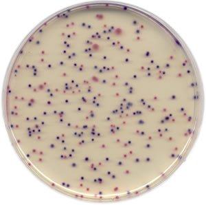
Here's how it works. You take a certain volume of your cell suspension - for the sake of simplicity, let's choose a volume of 1 ml - and disperse the suspension onto some sort of nutrient-containing plate. In our work, we use polystyrene petri dishes with a nutrient-enriched agarose. The cell suspension spreads out over the agar and the bacteria stick to the surface, start feeding on the nutrients in the agar, and begin to divide and proliferate. As the cells reproduce, they do not move away from each other. Instead, they sort of remain stuck together. And as they continue to proliferate, they create a little cell colony. After a certain incubation time, say 24 hours, each viable cell on the plate gives rise to its own discreet colony. Then you go through and count the number of colonies on the plate. Let's say for example that you count 100 colonies on a given plate. Since you started with a volume of 1 ml, that means that your concentration is 100 CFU/ml, where CFU = colony forming unit.

But there's a bit of a problem with my example, because 100 colonies on a plate is not a realistic number. More likely, the number of colonies would be several-fold larger; possibly in the thousands. That's a lot of counting...1 colony, 2 colonies, 3 colonies, 4...

There are ways to make this process a bit easier. Probably the most obvious option would be to divide the plate into even sections. If you divide the plate into, say, 4 equal area segments, then you only need to count the number of cells in one of those segments and simply multiply that number by 4. Either way, though, it's not a lot of fun and I'm not looking forward to it.